


The Discovery of X-Rays
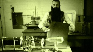
In late 1895, a German physicist, W. C. Roentgen was working with a cathode ray tube in his laboratory. Working with tubes similar to our fluorescent light bulbs, he evacuated the tube of all air, filled it with a special gas, and passed a high electric voltage through it. When he did this, the tube would produce a fluorescent glow.
Roentgen shielded the tube with heavy black paper, and found that a green coloured fluorescent light could be seen coming from a screen sitting a few feet away from the tube. He realised that he had produced a previously unknown “invisible light,” or ray that was being emitted from the tube; a ray that was capable of passing through the heavy paper covering the tube. Through additional experiments he also found that the new ray would pass through most substances, casting shadows of solid objects on pieces of film. He named the new ray X-ray, because in mathematics “X” is used to indicate the unknown quantity.
In his discovery Roentgen found that the X-ray would pass through the tissue of humans, leaving the bones and metals visible. One of Roentgen’s first experiments was a film of his wife’s hand with a ring on her finger. He later realised that a number of objects could be penetrated by these rays, and that the projected image of his own hand showed a contrast between the opaque bones and the translucent flesh. He later used a photographic plate instead of a screen, and an image was captured. In this way an extraordinary discovery had been made; that the internal structures of the body could be made visible without the necessity of surgery.
The invention of the x-ray created an amazing step forward in the history of medicine. For the first time ever, the inner workings of the body could be made visible without having to cut into the flesh.
The news of Roentgen’s discovery spread quickly throughout the world. Scientists everywhere could duplicate his experiment because the cathode tube was very well known during this period. In early 1896, X-rays were being utilised clinically in the United States for things such as bone fractures and gunshot wounds.
By 1896 an x-ray department had been set up at the Glasgow Royal Infirmary, one of the first radiology departments in the world. The head of the department, Dr John Macintyre, produced a number of remarkable x-rays; the first x-ray of a kidney stone; an x-ray showing a penny in the throat of a child, and an image of a frog’s legs in motion. In the same year Dr Hall-Edwards became one of the first people to use an x-ray to make a diagnosis – he discovered a needle embedded in a woman’s hand. In the first twenty years following Roentgen’s discovery, x-rays were used to treat soldiers fighting in the Boer war and those fighting in WWI, finding bone fractures and imbedded bullets. Much excitement surrounded the new technology, and x-ray machines started to appear as a wondrous curiosity in theatrical shows.
It was eventually recognised that frequent exposure to x-rays could be harmful, and today special measures are taken to protect the patient and doctor. By the early 1900’s the damaging qualities of x-rays were shown to be very powerful in fighting cancers and skin diseases.
In 1901, Roentgen received the first ever Nobel Prize in Physics. This was a true acknowledgement of his remarkable discovery which was going to be highly beneficial for mankind in the coming years. His discovery revolutionised the entire medical profession and set the foundation for diagnostic radiology.
Wilhelm Roentgen died on February 10, 1923 in Munich at the age of 77, and had his lab notes burned after his death.